The Dragonfly enables researchers to image biological samples in excellent quality at much higher-speeds than conventional confocal microscopy.
It can be used to answer a range of biological questions in both live cell cultures and fixed tissue samples. The dragonfly can produce excellent resolution imaging in 3 dimensions, allowing researchers to visually reconstruct tissue structure and cell-cell interactions. Its high speed means large areas of samples can be imaged quickly using the mechanical stage and intuitive software. The dragonfly can image at very high resolution meaning it’s possible to distinguish structures just 0.2um apart, or even closer by using integrated deconvolution and camera super-resolution techniques.
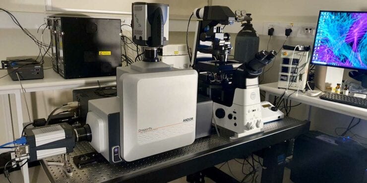
Optical configuration
CHIRI’s Dragonfly has 7 lasers channels from 405 to 730nm and matching emission filters, with the Borealis Perfect Illumination system to ensure stable and uniform excitation across the field of view. The Spinning Disk has 25um and 40um pinhole options to optimise for high-contrast imaging at a range of magnifications. The Nikon Ti2 base has 6 objectives corrected for confocal imaging including the 25x(NA1.05) Silicone-immersion objective ideal for live-cell imaging. The 60x oil-immersion objective can be combined with the 2x camera zoom lens for high-magnification acquisition at Nyquist sampling.
The two camera options allows users to optimise for high-resolution or high-sensitivity, including capacity for simultaneous imaging in 2 channels.
The Micropoint Photostimulation system provides a tool for photobleaching or ablation applications such as FRAP and FRET.
Accessories
- SRRF Stream – real-time camera super-resolution
- GPU accelerated deconvolution
- Tokai HIT stage top Incubator
- Perfect Focus System (PFS)
- Brightfield and Differential Interference Contrast (DIC)
- Laser-illuminated widefield epifluorescence function
- Bitplane Imaris software for visualisation and 3D rendering
User information
Prospective users must complete a comprehensive training program with facility staff, which requires about three hours.
For further information on the platform or bookings, please email Michael Nesbit at michael.nesbit@curtin.edu.au.
For enquiries about access to the CHIRI Lab facility, contact CHIRI Administration chiri@curtin.edu.au.